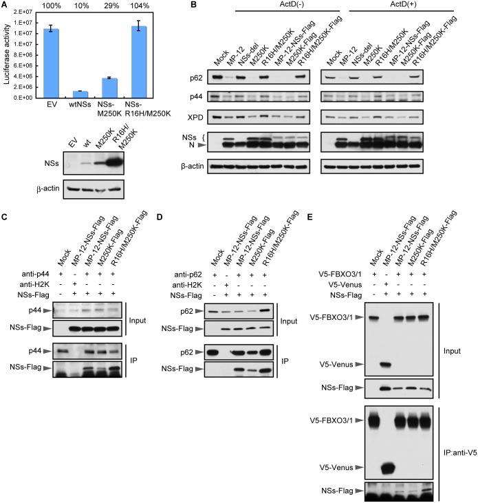
Fig 6. Effect of mutations in NSs on its interaction with TFIIH subunits.
(A) HeLa cells were co-transfected with NSs expression plasmid or empty vector (EV) along with reporter plasmid encoding Renilla luciferase. Luciferase activities (relative light units) were measured at 24h post transfection (top panel). Whole cell lysates were prepared from the transfected cells described above and subjected to Western blot analysis by using anti-MP-12 and anti-β-actin antibodies (bottom panel). (B) HeLa cells were mock infected (Mock) or infected with MP-12 or its mutants at an m.o.i. of 3 and harvested at 16 h p.i. Whole-cell lysates were subjected to Western blot analysis by using anti-p62 antibody and p44 antibody, anti-XPD antibody, anti-MP-12 antibody or anti-β-actin antibody as primary antibodies. (C) HeLa cells were mock infected (Mock) or infected with the MP-12 carrying Flag-tagged NSs or NSs or mutant MP-12 carrying Flag-tagged NSs mutant at an m.o.i. of 3. At 8 h p.i., the cells were lysed and subjected to immunoprecipitation by using anti-p44 antibody. Precipitates were subjected to Western blotting and analyzed by using anti-p44 antibody (third panel) and anti-Flag tag antibody (bottom). The top two panels show Western blot analysis of the input lysates by using anti-p44 antibody (top panel) and anti-Flag tag antibody (second panel). (D) HeLa cells were mock infected (Mock) or infected with the MP-12 carrying Flag-tagged NSs or NSs or mutant MP-12 carrying Flag-tagged NSs mutant at an m.o.i. of 3. At 5 h p.i., the cells were lysed and subjected to immunoprecipitation by using anti-p62 antibody. Precipitates were subjected to Western blotting and analyzed by anti-p62 antibody (third panel) and anti-Flag tag antibody (bottom). The top two panels show Western blot analysis of the input lysates using anti-p62 antibody (top panel) and anti-Flag tag antibody (second panel). (E) HeLa cells were transfected with the plasmid encoding V5-tagged full-length FBXO3 or V5-tagged Venus protein and infected with the MP-12 carrying Flag-tagged NSs or mutant MP-12 carrying Flag-tagged NSs mutant at 16 h post transfection. At 8 h p.i., the cells were lysed and subjected to immunoprecipitation using anti-V5 antibody. Precipitates were subjected to Western blotting analysis by using anti-V5 antibody (third panel) and anti-Flag tag antibody (bottom panel). The top two panels show Western blot analysis of the input lysates using anti-V5 antibody (top panel) and anti-Flag tag antibody (second panel).

