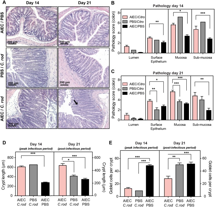
Fig 7. Mucosal epithelial restitution is delayed by AIEC following infectious colitis.
(A) H&E staining of colonic sections from C57BL/6 mice infected as indicated, on day 14 and day 21 after C. rodentium infection. Images are representative of 2 experiments with 5 mice per group per time point. Arrows indicate submucosa edema, desquamation, hyperplasia, inflammatory cellular infiltrates and epithelial sloughing. Original magnification 200x. Quantification of colonic pathology on day 14 (B) and day 21 (C) after C. rodentium infection. Measurements represent an average of at least 5 views per section and are the means with SEM from 5 mice. Quantification of crypt length (D) and goblet cell numbers (E) on day 14 and day 21 after C. rodentium infection. Measurements are from at least 5 views per section. Data are expressed as the means ±SEM of 5 mice per group/time point from 2 separate experiments. *p<0.05, **p<0.01, and ***p<0.001 (one way ANOVA with Tukey).

