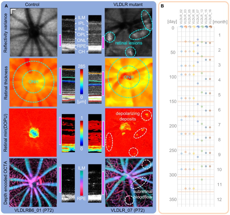
Fig 3. Multi-functional OCT images and timeline of measurements.

(A) Contrast specific projections were used for a quick identification of retinal abnormalities in the longitudinal investigation of the VLDLR mutant retina. An age-matched (P72) control mouse is compared to a Vldlr-/- mouse. Retinal lesions can be identified in reflectivity variance projections of the outer retina. Retinal thickness changes are present, especially at the location of lesion sites and is evaluated in a circumpapillary region excluding the optic nerve head (ONH). Depolarizing deposits within the retina of the Vldlr-/- mouse can be identified by a minimum DOPU projection. OCTA provides 3D contrast of the retinal microvasculature. Three plexuses can be visualized in the animals and pathological intraretinal neovascularization in the ONL was observed. (scale bar: 100 μm; pink bar: depth range which was used for the respective parameter’s en-face projection) (B) Image acquisition timeline representing the measured mutant mice at the respective postnatal days by a circle. For the evaluation process, the individual measurements were clustered per month.
