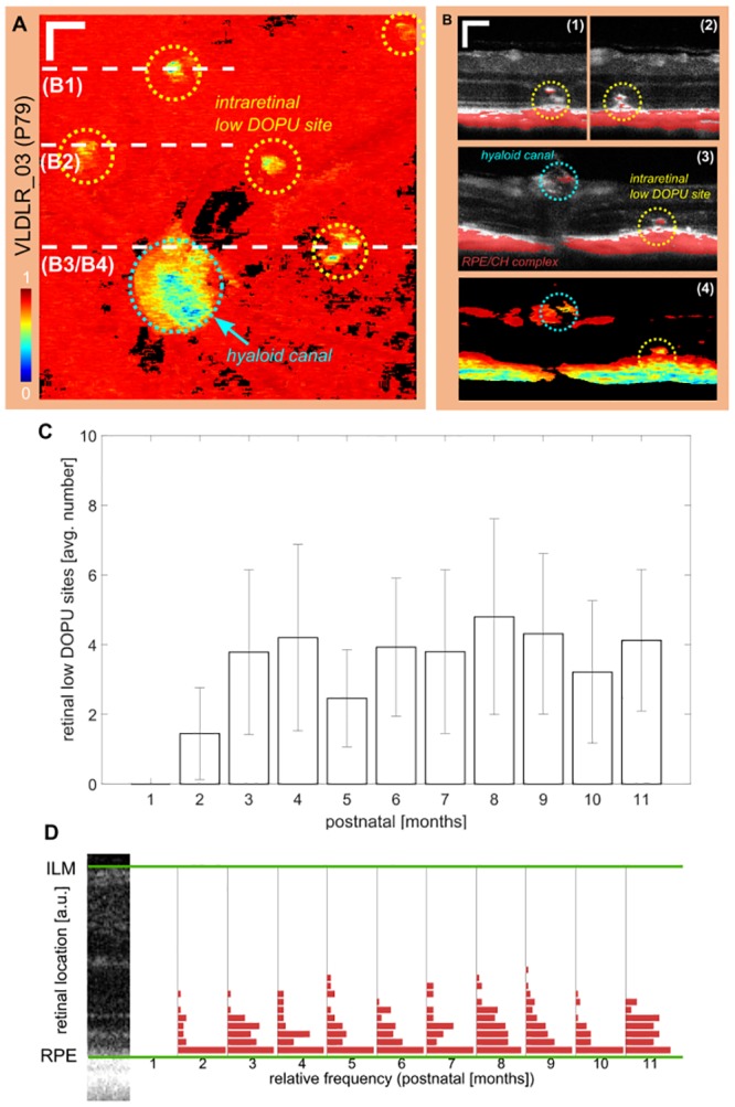
Fig 6. Intraretinal deposits of migrated melanin pigments.

(A) Intraretinal minimum DOPU projection of an exemplary mouse at P79. 6 clusters of low DOPU values can be identified. The biggest accumulation (cyan circle) is associated to melanin pigments at the hyaloid canal. The remaining sites are related to melanin migration during lesion development. (scale bar: 100 μm) (B) Exemplary reflectivity B-scans with a superimposed thresholded DOPU image (DOPU ≤ 0.5) at the lesion sites as shown in (A). Intraretinal deposits with low DOPU can clearly be identified along with disruption of the RPE in the DOPU B-scan of B3. (C) Mean number of low DOPU sites for all eyes clustered monthly. Error bars indicate the standard deviation. (D) The retinal location of the deposits was evaluated and binned into 20 equally spaced slabs between the RPE and the ILM. Most deposits are located close to the RPE and range up to the OPL.
