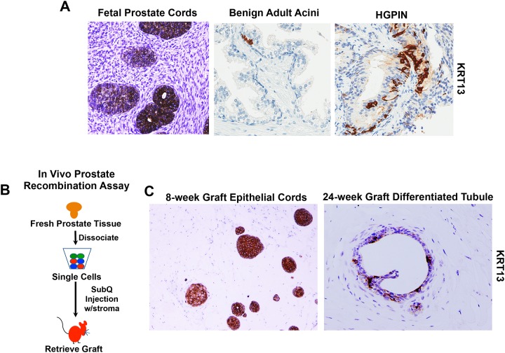
Fig 2. IHC analysis of KRT13 expression in fetal and adult prostate tissue and recombinant grafts.
(A) Immunohistochemical (IHC) analysis of fetal prostate tissue, benign adult prostate tissue, and HGPIN lesion. Representative images of fetal tissue 14–18 weeks gestation are shown. KRT13+ Benign and HGPIN lesions were routinely observed in radical prostatectomy specimens. (B) Schematic of in vivo prostate recombination assay. All grafts were generated from freshly dissociated prostate cells combined with hFPS and injected subcutaneously with Matrigel™ in vivo. (C) IHC analysis of recombinant grafts stained for KRT13. 8-week graft demonstrates predominantly cord-like structures, and 24-week grafts demonstrate differentiated tubules with lumens. Grafts generated from 3 individual prostate specimens were collected at each time period (<12 weeks and >24 weeks). Representative images of KRT13 expression are shown.

