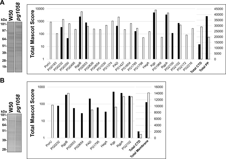
Fig 4. Aberrant localisation of T9SS substrates in the pg1058 mutant.
P. gingivalis W50 and pg1058 mutant periplasm (A) or total membrane (B) fractions were separated via SDS-PAGE and stained with SimplyBlue™ SafeStain. Each lane was divided into segments and analysed by LC-MS/MS. When the same protein was identified from multiple gel segments the Mascot scores were summed. The total Mascot score for the substrates identified from W50 (black bars) and the pg1058 mutant (white bars) were plotted on a logarithmic axis (left Y-axis). The total Mascot score for the combined substrates and the total Mascot scores for the combined periplasmic or membrane proteins were plotted on a linear axis (right Y-axis). Proteins are indicated by the Locus Tag in the P. gingivalis W83 strain unless previously designated with a protein ID.

