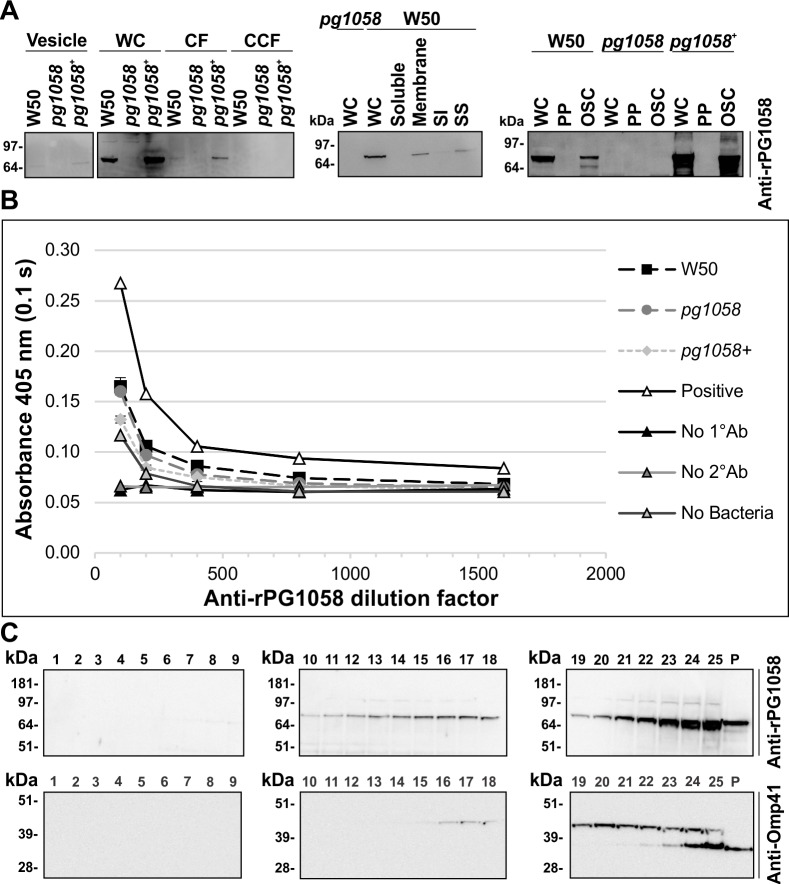
Fig 8. PG1058 is an OM-associated periplasmic protein.
A. P. gingivalis W50, pg1058 mutant and pg1058+ complement strain cultures were fractionated, separated via SDS-PAGE and immunoblotted on nitrocellulose membranes. The immunoblots have material derived from vesicles (5 x 10⁹ cells); whole-cells (WC; 2 x 10⁸ cells), culture fluid (CF; from culture containing 2 x 10⁹ cells) and vesicle-free cleared culture fluid (CCF; from culture containing 2 x 10⁹ cells); WC (2 x 10⁸ cells), soluble (5 x 10⁹ cells), total membrane (Membrane; 1.6 x 10¹⁰ cells), sarkosyl-insoluble membrane (SI; 1.6 x 10¹⁰ cells) and sarkosyl-soluble membrane (SS; 1.6 x 10¹⁰ cells) fractions; WC (5 x 10⁸ cells), periplasm (5 x 10⁸ cells) and osmotically-shocked cell (OSC; 5 x 10⁸ cells) fractions. Immunoblots were probed with anti-rPG1058 (1/10,000 dilution) and horse anti-mouse IgG-conjugated HRP secondary antibody (1/3,000 dilution). B. Whole-cell ELISA. P. gingivalis W50, pg1058 mutant, pg1058+ complement FKWC or lysed P. gingivalis W50 FKWC as a positive control (2 x 10⁹ cells per well) were probed with anti-rPG1058 antiserum (1/100, 1/200, 1/400, 1/800 and 1/1600 dilutions) followed by horse anti-mouse HRP-conjugated IgG secondary antibody (1/3,000) and developed with ABTS chromogenic substrate. The chromogenic reaction was suspended and detected at 405 nm. Other controls included no primary antibody control (No 1°Ab), no secondary antibody control (No 2°Ab) and no cell control (No Bacteria). Mean ± SEM, N = 3 for each strain except where only one replicate (N = 1) of the positive control (Positive) was assessed. C. P. gingivalis W50 total membrane was separated via sucrose density gradient centrifugation then fractions were separated by SDS-PAGE and immunoblotted on nitrocellulose membrane. Fraction numbers are indicated above the lanes and P refers to the pellet (unequal loading). Immunoblots were probed with anti-rPG1058 (1/10,000 dilution) or anti-Omp41 (1/10,000 dilution) followed by horse anti-mouse IgG-conjugated HRP secondary antibody (1/3,000 dilution).

