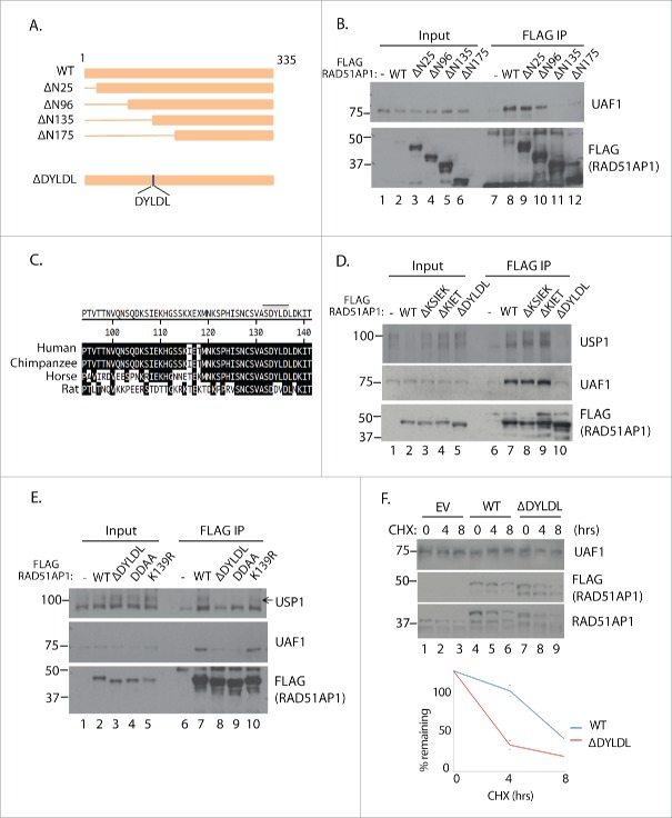
Figure 4.
Mapping of the UAF1-interacton region of RAD51AP1. A. Schematic diagram of the RAD51AP1 truncates used for analysis. The DYLDL sequence whose deletion abrogated the UAF1 interaction is highlighted below. B. 293T cells were transfected with the indicated plasmids, followed by anti-FLAG IP and anti-UAF1 western blot. C. Sequence alignment of the putative interaction area of RAD51AP. D, E. 293T cells were transfected with the indicated plasmids, followed by anti-FLAG IP and anti-UAF1 western blot. F. ∼24 hours after the HeLa cells were transfected with the plasmids, cells were treated with 10 uM cycloheximide for indicated time, then harvested for western blots. Below is the quantification of the anti-FLAG bands intensity from triplicate experiments.

