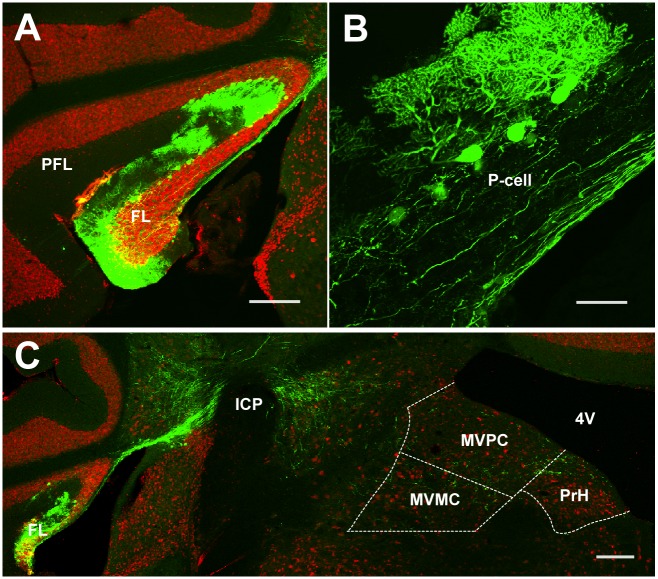
Fig 1. Anterogradely labeled FL P-cell axons in the medial vestibular nucleus (MVN) and prepositus hypoglossi (PrH) in case (#) 94R.
A, GFP expression in FL at 3 weeks after lentiviral injection. Sections were stained with anti-GFP (green) and neuronal marker anti-NeuN (red) antibodies. B, GFP was preferentially expressed in FL P-cells. C, GFP-labeled axons of FL P-cells in the ventromedial MVN. FL, flocculus; icp, inferior cerebellar peduncle; MVMC, magnocellular MVN; MVPC, parvocellular MVN; P-cell, Purkinje cell; PFL, paraflocculus; PrH, prepositus hypoglossi nucleus; 4V, fourth ventricle. Scale bars, 200 μm (A and C) and 50 μm (B).

