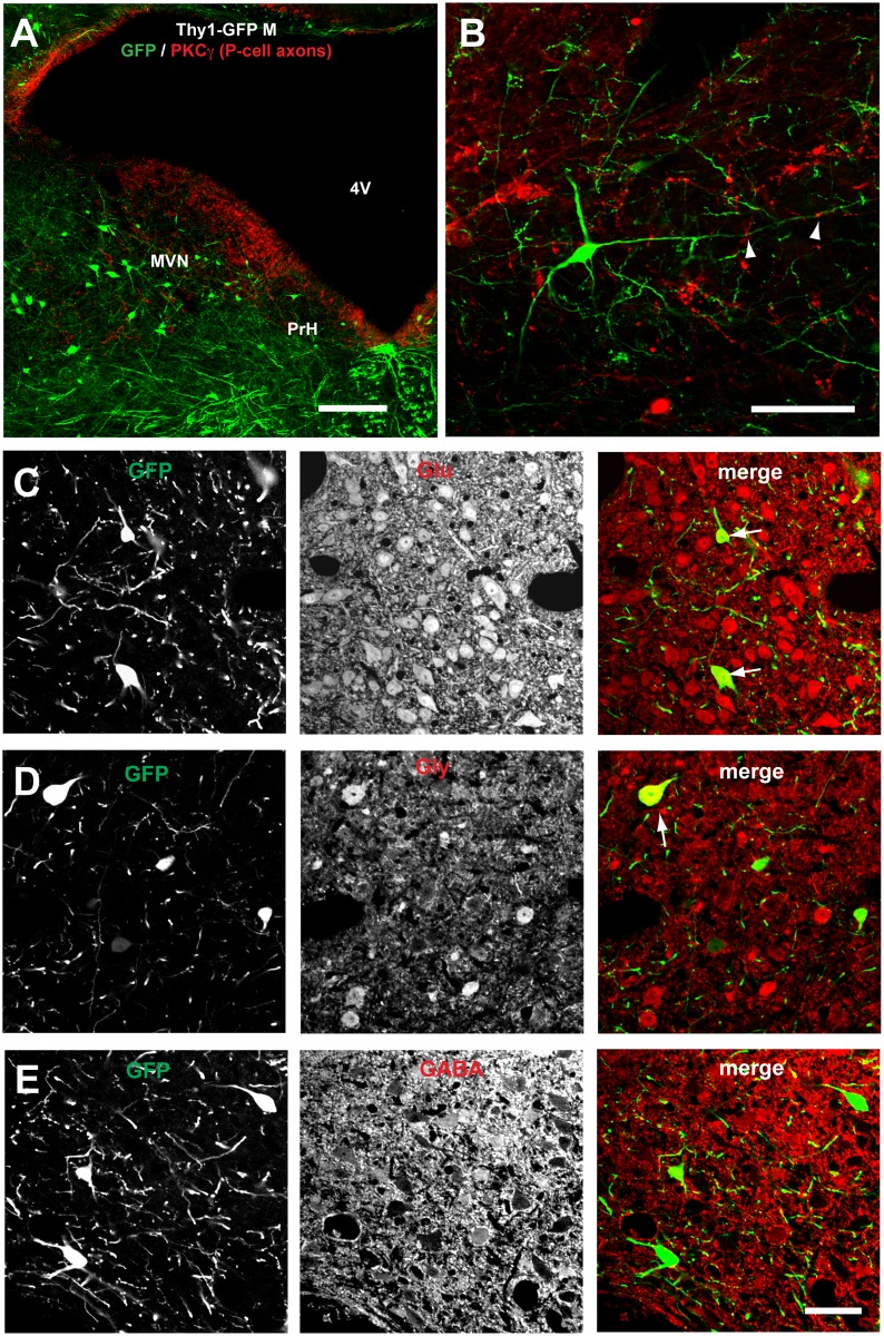
Fig 4. Neurons that receive FL P-cell projections on their distal dendrites in the parvocellular MVN/PrH are predominantly glutamatergic.
A and B, Representative images of MVN/PrH neurons of Thy1-GFP M-line transgenic mouse that were stained with anti-GFP (green) and anti-PKCγ (red) antibodies. C, D, and E, Images of coronal sections of MVN/PrH stained with either one of three antibodies against neurotransmitters (anti-Glu, anti-Gly, and anti-GABA). The majority of GFP-labeled neurons were stained positive for anti-Glu antibody (C), and some of the remaining neurons were positive for anti-Gly antibody (D), but not positive for anti-GABA antibody (E). Scale bars, 200 μm (A), 100 μm (B), and 50 μm (C–E). GABA, γ-aminobutyric acid; Glu, glutamate; Gly, glycine.

