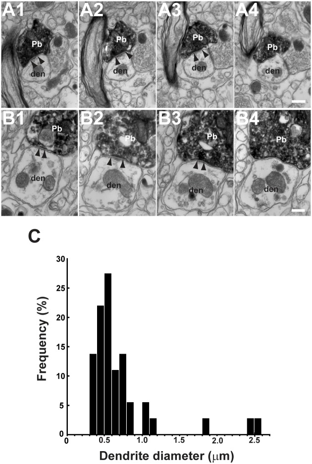
Fig 5. Axodendritic synapses between FL P-cells and parvocellular MVN/PrH neurons.
A1–A4, B1–B4, Photographs of two examples of EM serial sections (interval, 0.14 μm) for FL P-cell axonal boutons forming symmetrical synapses on parvocellular MVN/PrH neurons. Edges of synapses were shown by arrowheads. C, Distribution of the mean diameters for post-synaptic dendrites. Note that FL P-cell axonal boutons formed synapses on relatively small diameter dendrites (mean, 0.73 μm; n = 40 dendrites from three mice). den, dendrite of MVN/PrH neuron; Pb, P-cell axonal bouton. Scale bars, 0.2 μm.

