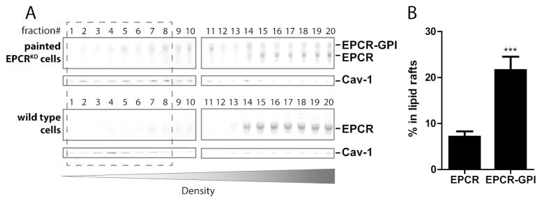
Figure 5. Localization of painted EPCR-GPI in lipid rafts.
To determine the localization of painted EPCR-GPI on cells, cell membranes were fractionated on a density gradient. (A) Distribution of endogenous EPCR and painted EPCR-GPI in membrane fractions of wild type EA.hy926 cells or EA.hy926 EPCRKD cells painted with 20 μg/ml EPCR-GPI. Caveolin-1 (cav-1) was used as a marker for lipid rafts. Representative blots of three independent experiments are shown. (B) Quantification of endogenous EPCR or EPCR-GPI in caveolin-enriched lipid raft (fractions 1–8) for wild type EA.hy926 cells (EPCR) and painted EA.hy926 EPCRKD cells (EPCR-GPI). Shown are mean ± SD of three independent experiments. *** P < 0.001.

