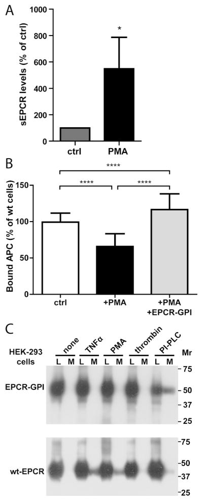
Figure 7. EPCR-GPI-mediated reconstitution of APC binding after PMA-induced EPCR shedding.

The ability of EPCR-GPI to restore APC binding after EPCR shedding was determined. (A) Shedding of EPCR was induced by treating wild type EA.hy926 cells with 0.2 μM PMA, and soluble EPCR (sEPCR) shed in the supernatant was measured by immunoprecipitation. Spontaneous shedding from untreated wild type cells was set to 100% (ctrl) (B) Reconstitution of APC binding by EPCR-GPI to the cell surface of PMA-treated cells was quantified using an on-cell Western assay. PMA-treated cells were incubation with 80 μg/ml EPCR-GPI (+PMA+EPCR-GPI) or left untreated (+PMA) before addition of APC. APC binding to non-treated wild type cells (ctrl) was used as 100%. Shown are mean ± SD of three independent experiments. * P < 0.05, **** P < 0.0001. (C) Sensitivity of EPCR-GPI to inflammatory mediator-induced shedding. HEK-293 cells transfected with EPCR-GPI or wt-EPCR were incubated with TNFα (10 ng/ml) for 12 hours or PMA (100 nM), thrombin (20 nM), or PI-PLC (0.5 U/ml) for 2 hours. Cell lysate (L) and conditioned medium (M) were analyzed for cellular and soluble shed EPCR by Western Blot using a goat anti-EPCR antibody. Shown is a representative example of two independent experiments.
