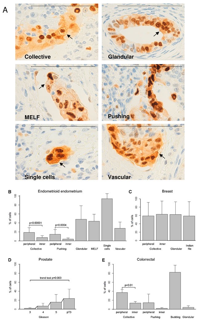
Figure 1. Membranous-cytoplasmic Ccnd1 expression at the invasive front is higher in peripheral cells, in large invasive cell clusters or in specific types of invasion.

A. Representative images showing Ccnd1 expression in endometrioid carcinomas of the endometrium (100μm bar). Different types of invasion are considered (collective, pushing, MELF, glandular, single cells/small cluster of cells, and vascular). Arrows indicate Ccnd1 stain in the membrane. Evaluation of the differences in membranous-cytoplasmic Ccnd1 expression among the different types of invasion in endometrioid endometrial carcinomas B., ductal breast carcinoma C., prostatic carcinoma according to Gleason grade or invasion beyond the prostate (pT3) D. and colonic carcinoma E. Bars represent mean percentages of positivity and segments one standard deviation. Significant differences between selected pairs are shown with their corresponding p-value, as computed with the linear mixed models. For prostate, p-value to evaluate the increasing trend is shown.
