Figure 1.

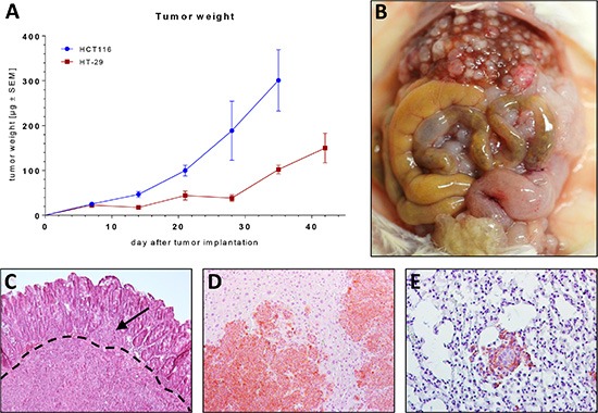
(A) Growth kinetics of orthotopically growing HT-29 and HCT116 tumor xenografts in NSG mice. (B) Situs of a NSG mouse bearing a HCT116 xenograft 35 days after tumor inoculation. (C) H & E section of a primary tumor (HCT116). Note the tumor bulk infiltrating the basement membrane (dashed line) and a tumor deposit on the luminal side of the basement membrane (arrow). (D–E) H & E section with immunohistochemical EpCAM staining (brown) of a liver metastasis (D) and a lung metastasis (E).
