
Figure 5. Activation of STAT3 signaling pathway was involved in the regulation of LGR5 expression.
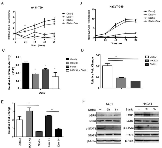
The MTT assay was performed to assess cell viability in A431 cells A. and HaCaT cells B. that were stably that were stably transfected with an inducible Tet-on IKKα shRNAs and the cells were treated with Stattic. C.. A luciferase reporter assay was carried out to evaluate LGR5 promoter activity in 293 cells after the treatment of IKKi-II and/or Stattic. All promoter luciferase intensity was normalized to the pRL renilla luciferase control reporter. * p < 0.05, ** p < 0.01. D.. ChIP analysis in HaCaT cells and chemicals treatment indicated was performed to detect p-STAT3 binding to LGR5 gene. E.. ChIP analysis in A431 cells after Tet-on shIKKα transfected and chemicals treatment indicated was performed to detect p-STAT3 binding to LGR5 promoter gene. F.. A431 (Left) and HaCaT (Right) with treatment of Stattic were examined for the expression of selected proteins by Western analysis. ** p < 0.01