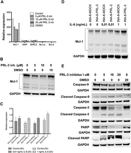
Figure 5. PRL-3 increases Mcl-1 expression, and PRL-3 inhibition reduces Mcl-1 expression and induces activation of the intrinsic apoptotic pathway.

(A) Expression of mRNA of the anti-apoptotic genes Bcl-xL, Bcl-2, Mcl-1, XIAP and BIRC2 in INA-6-WT cells. Cells incubated for 24 hours with IL-6 (1 ng/mL) in 10% FBS in RPMI and increasing concentrations of PRL-3 inhibitor I. (B) Expression of Mcl-1 protein in human myeloma cell line INA-6-WT after incubation without or with 5 or 10 μM of PRL-3 inhibitor I for 24 hours. (C) Expression of Mcl-1 mRNA in INA-6-MOCK and INA-PRL-3, measured at start of experiment (0 h) and after 3 hours without or with 0,01 ng/mL or 1 ng/mL IL-6, respectively. (D) Expression of Mcl-1 protein in INA-6-MOCK and INA-6-PRL-3 after 3 hours without or with 0,01 ng/mL or 1 ng/mL IL-6, respectively. (E) Expression of Caspase-9, cleaved Caspase-9, Caspase-3, cleaved Caspase-3, PARP and cleaved PARP in human myeloma cell line INA-6-WT after treatment without or with 5, 10 or 20 μM of PRL-3 inhibitor I or DMSO for 24 hours. GAPDH was used as a loading control.
