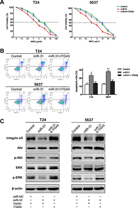
Figure 5. MiR-31/ITGA5 axis increases sensitivity of UBC cells to MMC via inactivating Akt and ERK pathways.

(A) Following 24 h treatment with MMC at different concentrations, MTT assay showed the survival rates of UBC cells co-transfected with miR-31 and empty plasmid were remarkably lower than those of cells co-transfected with miR-NC and empty plasmid (control); while co-transfection of miR-31 and ITGA5-expressing plasmid abrogated this effect. (B) Representative pictures of cell apoptosis distribution as detected by FCM analysis; region Q1, Q2, Q3 and Q4 shows dead, late apoptotic, early apoptotic and living cells, respectively; Q2 and Q3 are collectively regarded as apoptotic cells (left panel). Co-transfection of ITGA5-expressing plasmid basically eliminated the significant aggravation of MMC-induced apoptosis arising from miR-31 expression (right panel). (C) As indicated by western blot assay, overexpression of miR-31 significantly reduced the protein levels of ITGA5, phospho-Akt and phospho-ERK, while both Akt and ERK were reactivated again after ITGA5 expression was restored. Data are presented as mean ± SEM; *P < 0.05; **P < 0.01.
