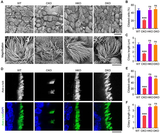
Figure 3. Cyld/Hdac6 DKO mice are protected from ciliary defects in the tracheal epithelium.

A. Scanning electron microscopy images of cilia in WT, CKO, HKO, and DKO mouse tracheal epithelia. Scale bars, 3 μm. B. and C. Experiments were performed as in A, and the percentage of ciliated cells (B) and ciliary length (C) were quantified. D. Immunofluorescence images of tracheal epithelial cilia in WT, CKO, HKO, and DKO mice, stained with acetylated α-tubulin (ace-α-tub) antibody and DAPI. Scale bar, 5 μm. E. and F. Experiments were performed as in D, and the percentage of ciliated cells (E) and ciliary length (F) were quantified. ***P < 0.001; ns, not significant. Data are represented as mean ± SEM.
