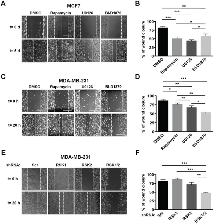
Figure 2. RSKs control migration of MDA-MB-231 cells.

A. MCF7 cells were subjected to wound healing assays in 1% FBS media containing vehicle (DMSO), rapamycin (20 nM), U0126 (10 μM), or BI-D1870 (10 μM). Media were replaced with fresh media containing vehicle or inhibitors daily. Representative photographs at the indicated times from three independent experiments performed in triplicate are shown. Magnification: x10. B. Percentage of wound recovery was determined in the experiments shown in A as described in Materials and Methods, and results were quantified as means ± SEM. (*p<0.05; **p<0.01; ***p<0.001). C. MDA-MB-231 cells were subjected to wound healing assays as described in A. D. Percentage of wound recovery was determined in the experiments shown in C, and results were quantified as in B. E. Control (Scr), RSK1-, RSK-2, or RSK1/2-silenced MDA-MB-231 cells were subjected to wound healing assays in 1% FBS media. F. Percentage of wound recovery was determined in the experiments shown in E, and results were quantified as in B.
