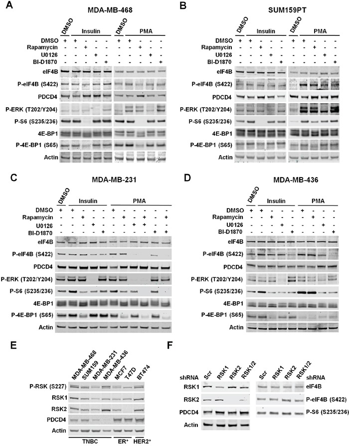
Figure 4. RSKs regulate phosphorylation of eIF4B and the levels of PDCD4 in TNBC cells with up-regulated MAPK pathway.

A. MDA-MB-468 cells were deprived of serum for 24 h, then treated with vehicle (DMSO), rapamycin (20 nM), U0126 (10 μM), or BI-D1870 (10 μM) for 30 min, followed by stimulation with insulin (100 nM) or PMA (50 ng/ml) for 2 h. Indicated proteins were analyzed by immunoblots. B. Serum-starved SUM159PT cells were treated as described in A. Indicated proteins were analyzed by immunoblots. C. MDA-MB-231 cells were deprived of serum for 24 h and then treated as described in A. Indicated proteins were analyzed by immunoblots. D. Serum-starved MDA-MB-436 cells were treated as described in A. Indicated proteins were analyzed by immunoblots. E. Cells were serum-starved for 24 h followed by stimulation with PMA for 2 h. Whole-cell extracts were resolved by SDS-PAGE and indicated proteins detected by immunoblot analysis. F. MDA-MB-231 cells were infected with lentiviruses expressing shRNAs targeted against a scrambled sequence (Scr), RSK1, RSK2, or RSK1/2. After selection, cells were serum-starved for 24 h followed by stimulation with PMA (50 ng/mL) for 4 h. Cell extracts were resolved by SDS-PAGE, and indicated proteins were analyzed by immunoblotting with specific antibodies.
