
Figure 4. Signal transduction system activation is involved in leptin-induced invasion in anaplastic thyroid cancer cells.
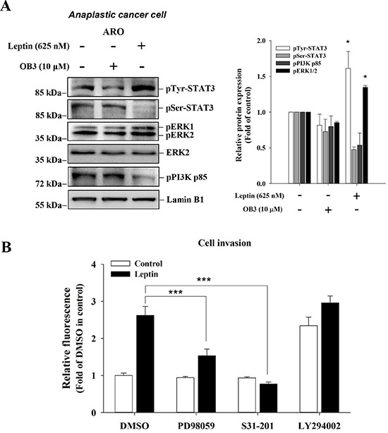
(A) Anaplastic thyroid cancer cells were incubated with 0.1 or 10 μM OB3, or leptin for 30 min. Nuclear extracts were prepared, proteins separated by PAGE, then immunoblotted with anti-phosphorylated MAPK (pERK1/2), anti-phospho-PI-3K p85, pSer-STAT3 antibody, and pTyr-STAT3 antibody. β-Actin was used as an internal loading control. Immunoblots were visualized by enhanced chemiluminescence (Amersham Life Science, Arlington Heights, IL, USA) and digital imaging. Results represent the mean ± SEM of three independent experiments. *p < 0.05 compared to untreated control. (B) Anaplastic thyroid cancer cells (1 × 10⁵/well) were starved in 0.1% serum-containing medium with 0.625 μM leptin with or without PD98059 (10 μM), LY294002 or STAT3 inhibitor for one hr, and then incubated in the absence or presence of 10 μM leptin at 37°C for 4 h and seeded into the upper chamber of the transwell using the Millipore system for cell migration, respectively. After 6 h, the cells were subjected to chemoattraction and migrated into the lower chamber. A fluorimetric detection system (Millipore) quantitated movement. Data were expressed as mean ± S.D. in triplicate. (***p < 0.001, were compared with DMSO).