
Figure 5. Effect of OB3 and leptin peptides on integrin αvβ3 gene expression, FAK activation and adhesion in thyroid cancer cell lines.
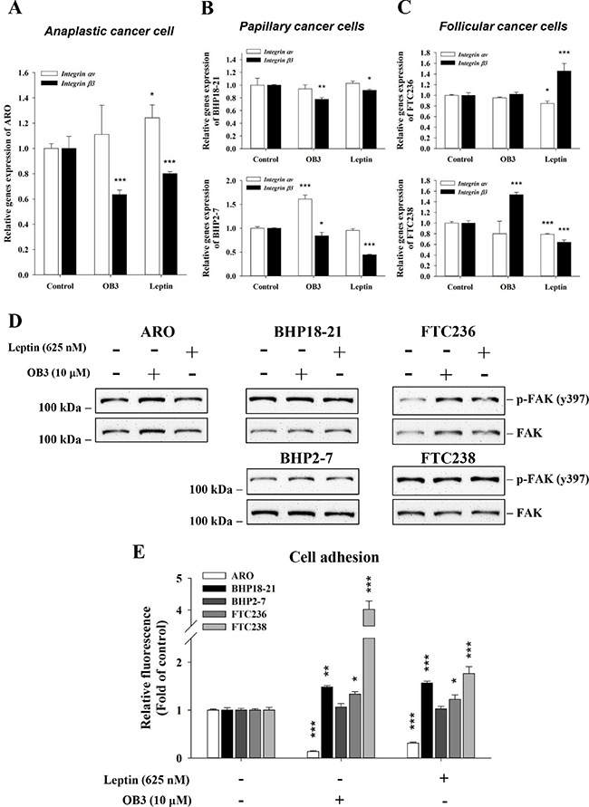
(A) Anaplastic, (B) papillary and (C) follicular thyroid cancer cells were treated with either leptin or OB3 for 24 h. Cells were harvested and total RNA was extracted. qPCR for integrin αv and β3 was conducted as described in the Materials and Methods. (D) Different thyroid cancer cells were incubated with 10 μM OB3, or 0.625 μM leptin for 30 min. Total cell extracts were prepared, proteins separated by PAGE, then immunoblotted with anti-phosphorylated FAK or total FAK antibody. β-Actin was used as an internal loading control. Immunoblots were visualized by enhanced chemiluminescence (Amersham Life Science, Arlington Heights, IL, USA) and digital imaging. (E) Thyroid cancer cells (5 × 10⁵/well) were starved in 0.1% serum-containing medium for 48 h. Cells were trypsinized and refed with medium with 0.625 μM leptin or 10 μM OB3 and cells were incubated at 37°C for 24 h. Cells were seeded into the 6-well cell culture plate for 6 h. The attachment cells were quantified using the fluorimetric detection system (Millipore). (Data were expressed as mean ± S.D. in triplicate. *p < 0.05, **p < 0.01, ***p < 0.001, were compared with control).