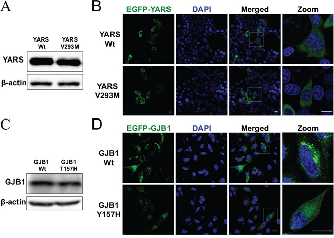
Figure 2. Functional analysis in cultured cells.

A. Western blot analysis of protein extracts obtained from HEK293 cells transfected with pFlag-YARS-Wt or pFlag-YARS-V293M vectors. The anti-Flag antibody was used to detect the interested band. B. Confocal images of HeLa cells transiently transfected with pEGFP-YARS-Wt or pEGFP-YARS-V293M vectors. Transfected cells were examined for green fluorescence 48 hours after transfection. Cell nuclei were stained with 4′-6-diamidino-2-phenylindole (DAPI; blue). Scale bar, 20 μm. C. Western blot analysis of GJB1 expression in HEK293 cells transfected with pEGFP-GJB1-Wt or pEGFP-GJB1-Y157H vector. The anti-GFP antibody was used to detected recombinant proteins. D. Confocal images of HeLa cells transiently transfected with pEGFP-GJB1-Wt or pEGFP-GJB1-Y157H constructs. Cells expressing wild type GJB1 displayed granules throughout the cytosol, whereas cells expressing the mutant GJB1 had diffuse intracellular staining. Scale bar, 20 μm.
