
Figure 2. Effects of metformin on bladder cancer proliferation and apoptosis.
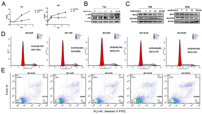
A. T24 and RT4 cells were seeded in 96-well plates with 0.5×10⁵ cells per well in growth media with or without metformin (20mM) and cultured for 5 days. Cell viabilities were estimated by MTT every other day. B. T24 cells were treated with different concentrations of metformin (0, 1, 5, 10 and 20 mM) for 24 h. The levels of total AMPK and phosphorylated AMPK were estimated by western blot assays. C. The effects of metformin on the levels of Bcl-2 and CyclinD1 were measured by western blotting when T24 and RT4 cells were treated with increasing concentrations of metformin. D. and E. T24 cells were treated with different concentrations of metformin and the numbers of cells at different stages of cycle were analyzed by flow cytometry (D), or stained with PI and FITC-labelled Annexin V and subsequently underwent flow cytometry analysis to determine the percentage of apoptotic cells (E).