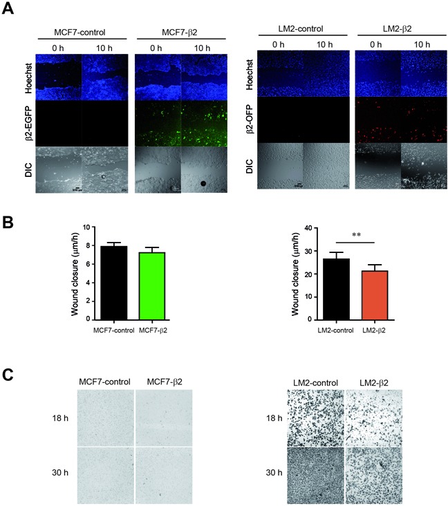
Figure 4. β2-chimaerin differentially affects cell migration and invasion in epithelial or mesenchymal-like cells.

A-B. MCF7-control, MCF7-β2, LM2-control and LM2-β2 cell lines were subjected to a wound healing assay. A. Representative images showing the wound closure at two different time points. Nuclei were stained with Hoechst to corroborate that equal number of cells were used in each case. B. Quantification of the relative migration of control versus β2-chimaerin expressing cells. Results are presented as means ± s.e.m (n=7) (**P < 0.01, unpaired two-tailed Student's t-test). C. Invasion of MCF7-control, MCF7-β2, and LM2-control and LM2-β2 cells was assessed using the Boyden chamber assay in the presence of Matrigel-coated filters. Images show crystal violet-stained migrated cells present at the lower face of the transwell membranes and are representative of one out of three different experiments.
