
Figure 4. Antitumor activity of Se-PFPs in HEY-xenografted nude mice.
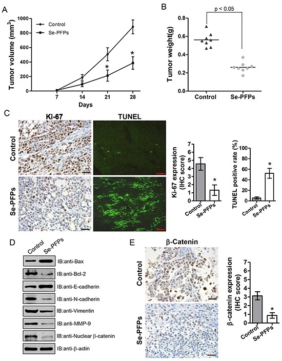
The volume A. and weight B. of tumors from HEY-xenografted nude mice were shown. HEY cells (1 × 10⁶) were injected subcutaneously into nude mice, and then administered with PBS or Se-PFPs (400 mg/day/kg weight) by gavage for 28 consecutive days. The tumor size was measured every 7 days. After 28 days of treatment, the mice were euthanized by CO2 inhalation. The tumors were separated and weighed. C. IHC staining for proliferation marker Ki-67 and TUNEL positivity, and scale bar represents 50 μm in Ki-67 staining and 500 μm in TUNEL staining, respectively. D. Western blot analysis was performed to determine the expression of Bax, Bcl-2, E-cadherin, N-cadherin, vimentin, MMP-9 and nuclear β-catenin in xenografted tumor tissues. β-actin was used as a control. E. IHC staining for nuclear β-catenin in tumors from HEY-xenografted nude mice. Scale bar = 50 μm. The statistic analysis was carried out by student's t-test. *P<0.05.