
Figure 3. Activation of Bax correlates with apoptosis induced by YY1 overexpression.
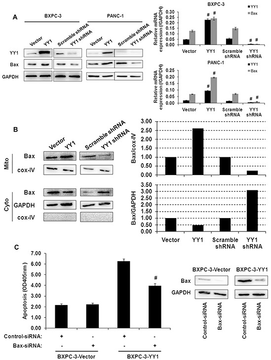
A. Bax expression in YY1 overexpressing or knockdown BXPC-3 and PANC-1 cells was measured by quantitative RT-PCR and Western blot. Results are representative of three independent experiments and are presented as the mean ± SD (bars). #p< 0.01. B. Bax expression in mitochondrial membrane and cytosolic fractions of BXPC-3 cells was measured by Western blot. Each blot was measured by densitometry (right panel) to assess the translocation of proteins. Results are representative of three independent experiments. C. YY1 overexpressing BXPC-3 cells were transfected with Bax siRNA for 48 h, after which the extent of apoptosis (left panel) and Bax protein expression levels (right panel) were measured. Results are representative of three independent experiments and are presented as the mean ± SD (bars). #p< 0.01.