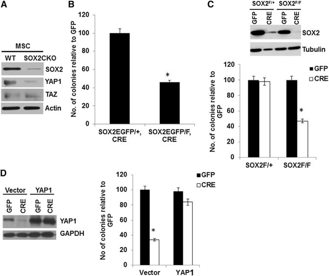
Figure 3. SOX2 Deletion in Primary BM-MSCs Leads to Decreased YAP1 and Reduced Colony Formation that Can Be Rescued by YAP1.

(A) Western analysis of SOX2, YAP1, and TAZ in BM-MSCs isolated from WT or SOX2 CKO mice.
(B) Colony assay (in fibroblast colony-forming units [cfu-f]) of BM-MSCs isolated from 4-week-old SOX2EGFP/+, osterix-CRE (heterozygous) or SOX2EGFP/F, osterix-CRE (SOX2 CKO) mice; 105 cells were plated in triplicate and analyzed as in Figure 2E. *p < 0.05.
(C) Western analysis and colony assay of SOX2F/+ or SOX2F/F BM-MSCs following in vitro CRE-mediated deletion of endogenous SOX2. Primary BM-MSCs were isolated from 4-week-old mice infected with control (GFP) or SOX2-deleting (CRE) virus. *p < 0.05.
(D) Rescue of the colony-forming ability of SOX2-deleted BM-MSCs by YAP1. Western analysis and colony assay were conducted on SOX2F/− BM-MSCs overexpressing YAP1 and depleted of endogenous SOX2 by CRE lentivirus infection.
*p < 0.05; error bars represent the average + SD. See also Figure S3.
