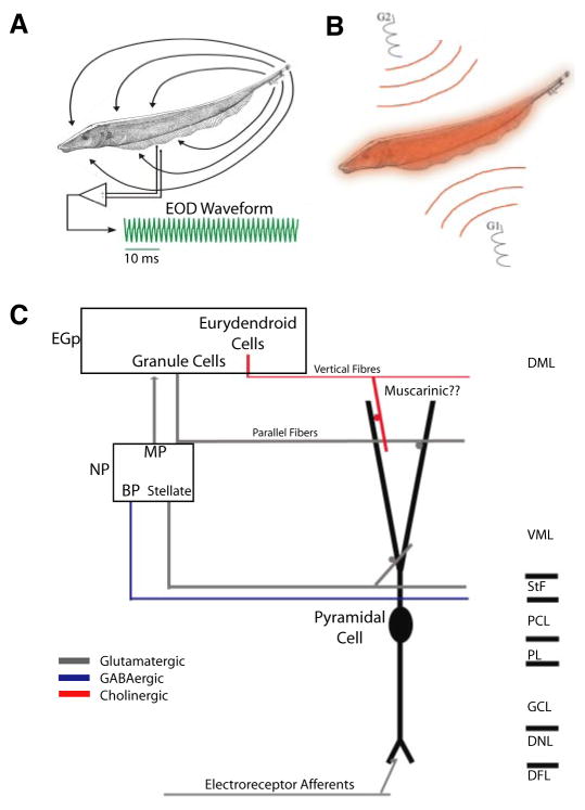
FIG. 1.
The electrosensory system. A: weakly electric fish generate an electric field around their body to electrolocate objects. For Apteronotus leptorhynchus, the electric organ discharge (EOD) waveform recorded at 1 point in space is quasi-sinusoidal with a frequency of 600–1,000 Hz (green trace). B: illustration of the Global stimulation geometry used. Amplitude modulations of the animals’ own EOD are delivered via 2 silver-silver-chloride electrodes (G1 and G2) located 19 cm away on each side of the animal. The perturbations of the EOD created are roughly spatially homogeneous on the animal’s skin surface. C: illustration of the relevant circuitry. Electroreceptor afferents on the animal’s skin detect amplitude modulations of the EOD and synapse unto pyramidal cells within the electrosensory lateral line lobe. Pyramidal cells project to the nucleus praeeminentialis (NP). Bipolar cells (BP) and stellate cells feed back, from the NP unto pyramidal cells by way of the tractus stratum fibrosum (StF). Multipolar cells (MP) project to granule cells (GC) within the eminentia granularis posterior (EGP) that feed back unto pyramidal cells via parallel fibers. Finally, pyramidal cells are thought to receive cholinergic muscarinic input from eurydendroid cells within the EGP via vertical fibers. DML, dorsal molecular layer; VML, ventral molecular layer; PCL, pyramidal cell layer; PL, plexiform layer; DNL, deep neuropil layer.

