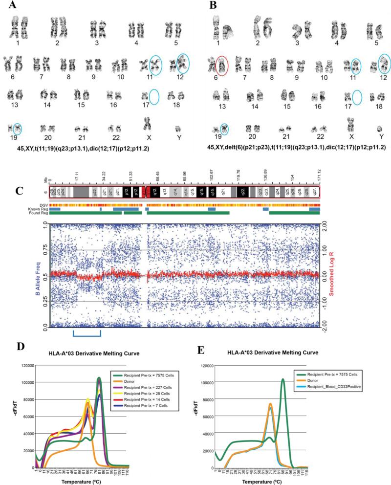
Figure 2. Assessment of patient-specific mismatched human leukocyte antigen (HLA) - haplotype loss using single nucleotide polymorphism array and melting curve analysis in recipient 2.
A) Karyotype of the diagnostic sample; B) Karyotype of the relapse sample showing a new finding of deletion in 6p; C) Single nucleotide polymorphism (SNP) array revealed the 15.9 Mb interstitial deletion (bracketed) that includes the HLA loci. The wide range of B allele frequencies (blue tracks) represent the presence of both patient and donor alleles; D) Melting curve analysis shows the ability to detect HLA-A*03 DNA in limited numbers of recipient pretransplant cells, but not in donor cells. -dF/dT represents derivative of fluorescence over temperature; E) Isolated leukemic CD45low CD33+ blasts from the recipient's peripheral blood and bone marrow show no detection of HLA-A*03 DNA, similar to that of the donor, suggesting loss of the recipient's mismatched HLA.

