
Figure 2.
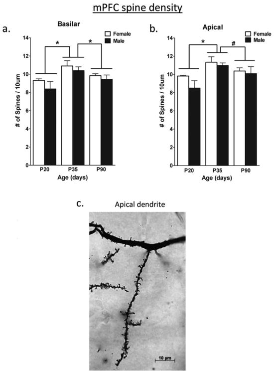
The density of dendritic spines at postnatal ages on layer V pyramidal neurons in the mPFC a) on the basilar dendrites and b) on the apical dendrites. Spines were pruned between P35 and P90. There were no sex differences. c) A photograph of dendritic spines visualized with a Golgi Cox stain. *p<.05; #p<.08 From Koss et al. (2014).