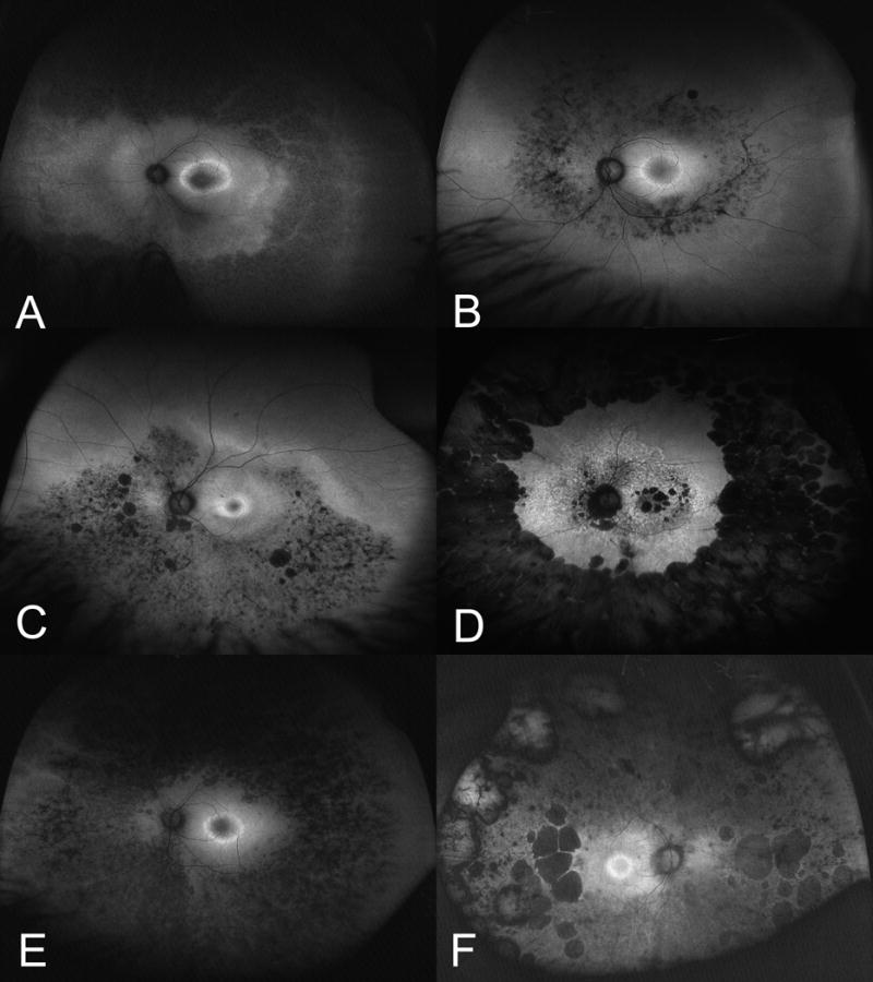
Figure 1. Ultra-widefield Fundus Autofluorescence Patterns in Retinitis Pigmentosa and Other Retinal Dystrophies.

A. Macular ring hyperautofluorescence with as seen in patients with RHO, USH2A, CEP290, RPGRIP1 and RPGR mutations. B. Ultra-widefield fundus autofluorescence in retinitis pigmentosa with USH2A mutation demonstrates diffuse macular hyperautofluorescence and a ring of hyperautofluorescence. C. Ultra-widefield fundus autofluorescence in retinitis pigmentosa with RHO or RPGR with hyperautofluorescence with patchy areas of hypoautofluorescence. D. Ultra-widefield fundus autofluorescence in retinitis pigmentosa with RDS mutations had a distinct pattern of diffuse peripheral hypoautofluorescence. E. Ultra-widefield fundus autofluorescence in retinitis pigmentosa with USH2A mutation had diffuse peripheral hypoautofluorescence. F. Ultra-widefield fundus autofluorescence in retinitis pigmentosa with PRPF31 or RHO mutations with associated optic nerve pallor appeared to have optic nerve hyperautofluorescence.
