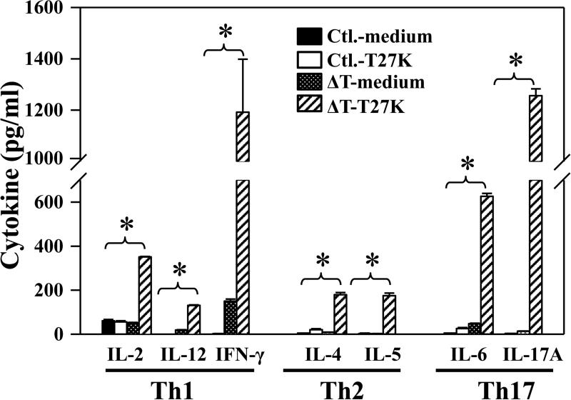
FIG. 3. Concentrations of representative Th1-, Th2-, and Th17-type cytokines detected in culture supernatants of splenocytes isolated from ΔT-vaccinated HLA-DR4 mice (ΔT) or nonvaccinated animals (Ctl.).
Ex vivo stimulation of both immune and naïve splenocytes was conducted using the T27K antigen. Cells incubated with medium alone served as a negative control. The asterisks indicate significantly higher concentrations of selected cytokines in vaccinated mice compared to non-vaccinated mice (P≤0.01). Each sample was analyzed in triplicate. The results are presented as mean values ± SEM.

