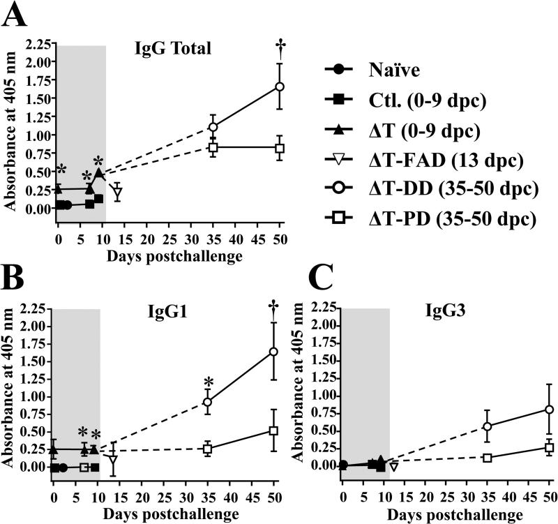
FIG. 6. Analysis of humoral immune response to Coccidioides infection revealed that the ΔT-vaccinated mice with disseminated disease had continuously rising antibody titers during the course of Coccidioides infection.
IgG total and subclass of IgG1, IgG2c and IgG3 responses to the T27K antigen by vaccinated (ΔT) and non-vaccinated mice (Ctl.) prior to and after challenge at 0, 7 and 9 dpc were determined by ELISA. IgG2c reactivity was not detected for all test samples. Sera obtained from ΔT-vaccinated mice with fatal acute disease (FAD) were also assayed at 13 dpc and for mice with pulmonary (PD) and disseminated disease (DD) at 35 and 50 dpc. Sera samples were diluted 1 to 50. The results are expressed as absorbance at 405 nm and presented as means ± SEM. The asterisks indicate statistically significant differences (P < 0.05) between vaccinated mice versus non-vaccinated control animals and the dagger indicates significant difference between the pulmonary versus disseminated groups.

