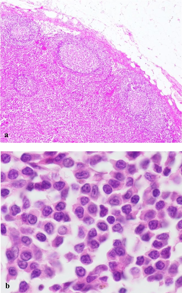
Fig. 3.
The histological features of canine TZL. (a) Neoplastic lymphocytes expand in the paracortex and are peripheralizing the fading lymphoid follicles. Hematoxylin and eosin (H&E) stain. × 40. (b) Neoplastic cells are small to intermediate in size. The nuclei are small and sometimes have sharp shallow indentations. Some nuclei have small central nucleoli. H&E stain. × 1,000.

