. 2016 Sep 28;46(5):601–609. doi: 10.4070/kcj.2016.46.5.601

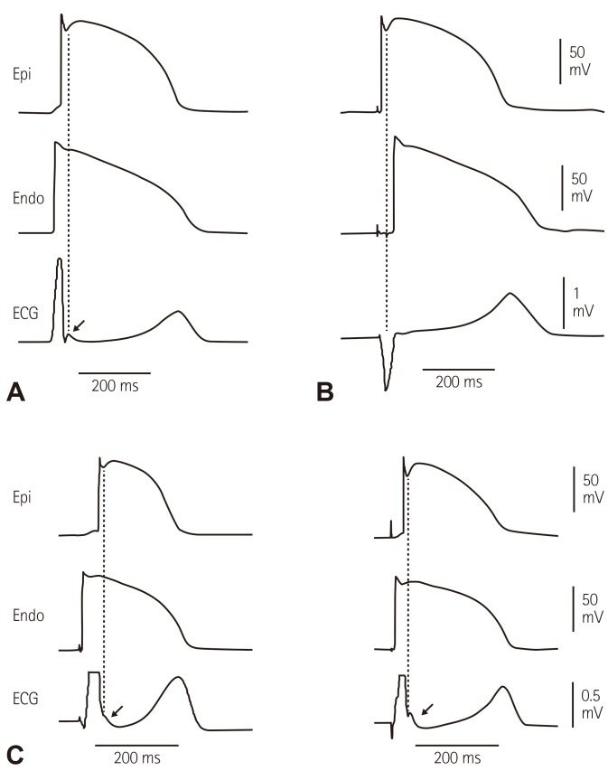
Fig. 1. The sequence of ventricular activation influences the appearance of J wave in a transmural ECG recorded from coronary-perfused canine left ventricular wedge preparation. (A) Stimulation of the endocardial (Endo) surface causes the epicardial (Epi) surface to be activated last. In such case the J wave is aligned with Ito-mediated epicardial AP notch. (B) Stimulation of the epicardial surface activates it before the endocardial surface. This causes the epicardial AP notch to occur simultaneously with the QRS, hiding the J wave. Reproduced from Yan and Antzelevitch4) with permission. (C) Endocardial activation at different locations can cause the J wave to occur at the end of the QRS, manifesting as slurred (left panel) or notched (right panel) QRS. Reproduced from Badri et al.5) with permission.

Fig. 1