. 2016 Sep 28;46(5):601–609. doi: 10.4070/kcj.2016.46.5.601

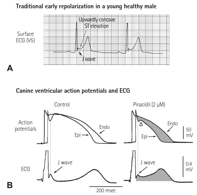
Fig. 3. Cellular basis for the traditional ER. (A) Surface ECG (lead V5) recorded from a 17-year-old healthy African American man. Note the presence of a small J wave and upwardly concave ST segment elevation. (B) Simultaneous recording of transmembrane action potentials from epicardial (Epi) and endocardial (Endo) regions and a transmural ECG in an isolated arterially perfused canine left ventricular wedge. A J wave in the transmural ECG is present due to the action potential notch in the epicardium but not the endocardium. Perfusion of the preparation with pinacidil (2 µmol/L), an ATP-sensitive potassium channel opener, causes partial loss of the action potential dome in the epicardium that separates earlier from the endocardium during phase 2 plateau phase, resulting in upwardly concave ST segment elevation in the ECG resembling the traditional ER. Reproduced from Hlaing et al.12) with permission. ECG: electrocardiography.

Fig. 3