. 2016 Sep 28;46(5):601–609. doi: 10.4070/kcj.2016.46.5.601

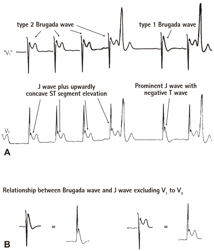
Fig. 6. Example to demonstrate the counterparts of type 1 and 2 Brugada waves. (A) The bottom ECG tracing exhibiting prominent J waves in a left precordial lead V5 was obtained from a 34 year-old Chinese man who survived from idiopathic VF. The J wave wad accentuated after a pause, which was accompanied by a negative T wave. The upper ECG tracing was produced by turning the QRS complex in the bottom ECG tracing (V5) by 180 degree to simulate a pseudo V1 tracing. The upper tracing was then evaluated by 27 electrophysiologists, among whom 24 agreed that there were type 2 and type 1 Brugada waves in this tracing. (B) Type 2 Brugada wave equates J wave plus upwardly concave ST segment elevation in leads excluding V1 to V3; type 1 Brugada wave is the counterpart of a prominent J wave with a negative T wave in leads excluding V1 to V3. ECG: electrocardiography.

Fig. 6