Fig. 5.

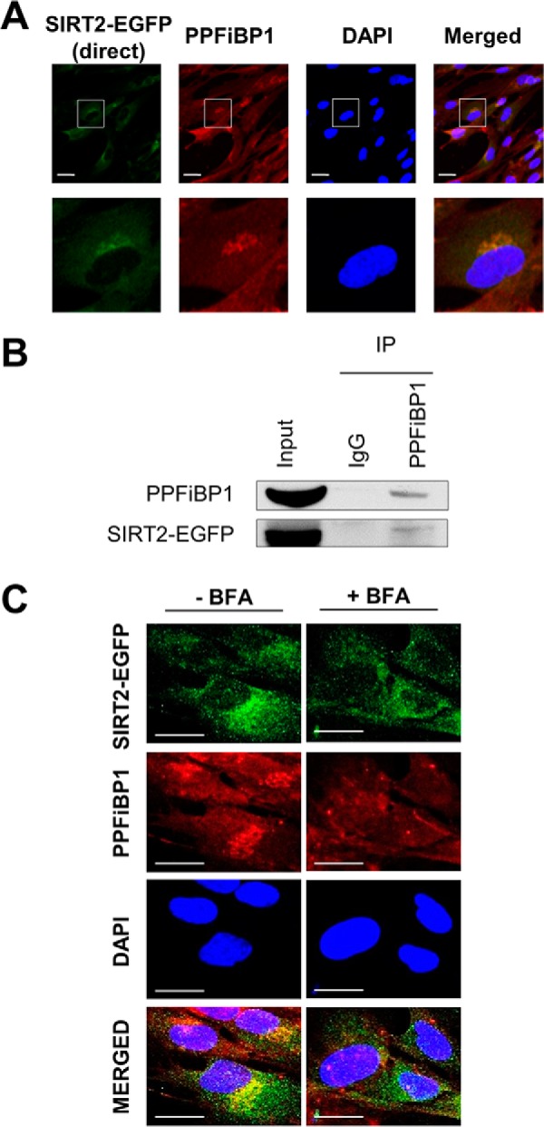
SIRT2 interacts and colocalizes with liprin-β1 (PPFiBP1). A, SIRT2 colocalizes with PPFiBP1. SIRT2-EGFP MRC5 cells were imaged by direct fluorescence (SIRT2-EGFP, green) or immunofluorescence microscopy (anti-PPFiBP1, red). Bars - 25 μm. B, PPFiBP1 reciprocally interacts with SIRT2. PPFiBP1 and IgG control IPs were performed in parallel. Western blotting analysis was done using anti-GFP antibody to detect SIRT2-EGFP. Input - 3% (v/v) of IP input fraction, IP - 30% (v/v) of IP elution fraction. C, PPFiBP1 localization was assessed with or without Brefeldin A treatment in MRC5 cells stably expressing SIRT2-EGFP. Immunofluorescence microscopy was performed using anti-EGFP (green) and anti-PPFiBP1 (red) antibodies. DAPI is a nuclear stain. Bars - 50 μm.
