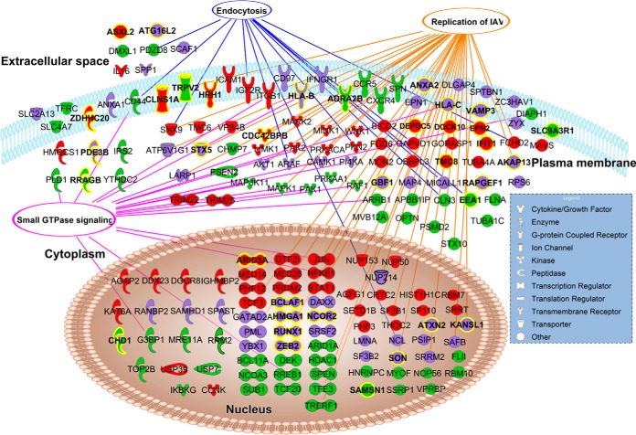
Fig. 2.
The phosphorylation profiles change upon influenza A virus infection for host proteins known to be involved in different stages of viral infection. In total 180 proteins of the 1113 uniquely phosphorylated proteins were directly associated with the biological function “viral infection” according to IPA®, and 177 of these had annotated subcellular locations. These 177 proteins are shown and their molecular type is indicated by specific shapes. The purple nodes are proteins, which have unique phosphorylation sites in both uninfected (control) and infected conditions. The red nodes are proteins, which have unique phosphorylation sites only after infection. The green nodes are proteins that have unique phosphorylation sites only in the control sample. Proteins associated with small GTPase signaling (connected with magenta colored edges), replication of influenza A virus (connected with orange colored edges), and endocytosis (connected with blue colored edges) are highlighted with edges connected to the respective biological function. Phosphoproteins with novel phosphorylation sites are highlighted in bold text coding and yellow colored outlining.

