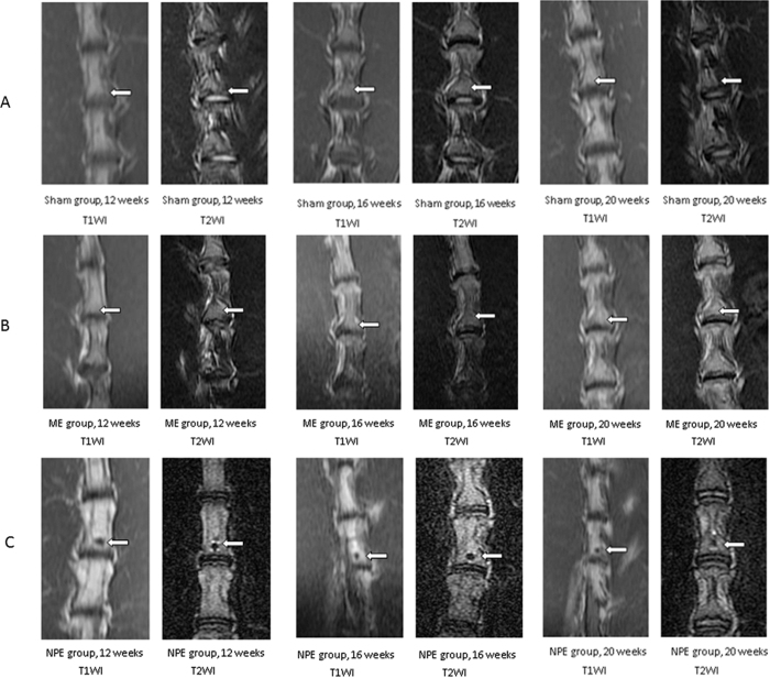
Figure 1.
(A) Representative serial MRI scans of the lumbar spine of a rabbit in three time points. In the sham group, no signal abnormality was observed in the picture. (B) The characteristic of vertebral body signal in the ME group was similar to the sham group. As time elapsed, there was no significant signal change in the embedment position. (C) In the NPE group, the hypointense signal in T1W and mixed signals with hypointense in T2W sequences were easily observed. From the 12-week time point to the 20-week time point, the sporadic hyperintense signals around the hypointense signal in T2W were diminished.

