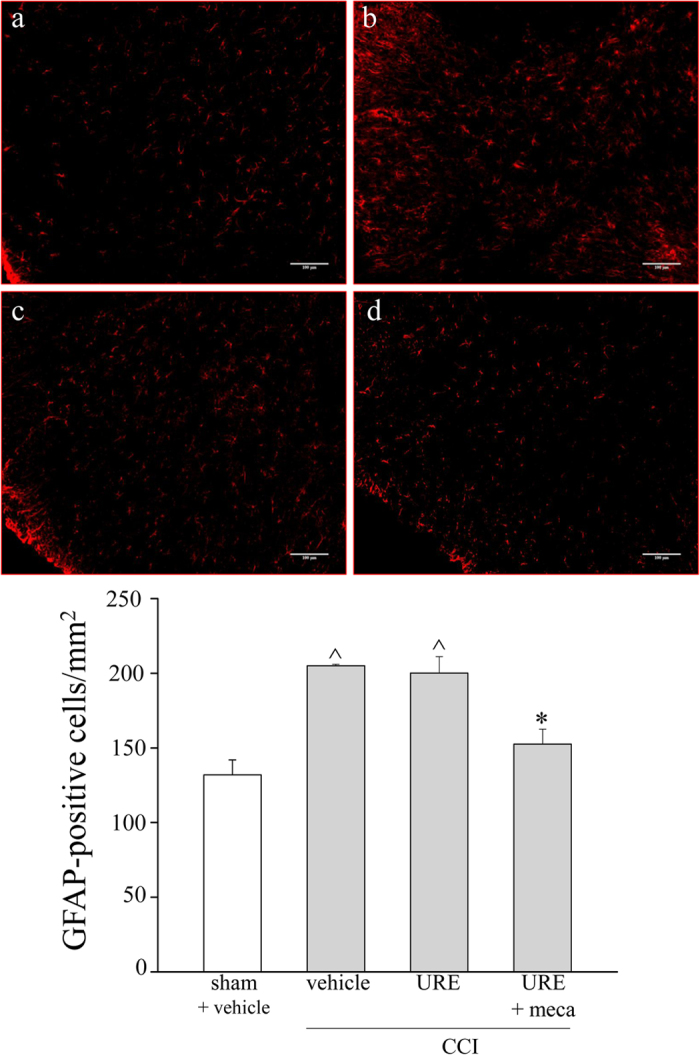
Figure 9. Glial profile in spinal cord scored with GFAP-positive cells 14 days after surgery.

URE (70 mg kg−1, p.o., daily) and mecamylamine (2.0 mg kg−1, i.p., bid) were administered starting on the day of surgery. The number of GFAP-positive cells was evaluated in the ipsilateral dorsal horn hemisection of the lumbar tract. In the upper panel, representative transverse sections of spinal cord imaged with 20X objective of (a) sham + vehicle; (b) CCI + vehicle; (c) CCI + URE; (d) CCI + URE + mecamylamine (meca). Quantitative analysis of cellular density was performed evaluating 6 animals for each group. Each value represents the mean ± SEM of 6 rats per group, performed in 2 different experimental sets. ^P < 0.05 versus sham + vehicle; *P < 0.05 versus CCI + vehicle.
