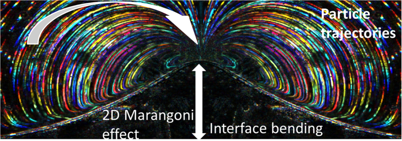
Figure 3. 2D Marangoni effect.
The composed microscopic dark field photograph of stable laser-induced whirl in pNA-1,4-dioxane solution in a thin layer. Multicolor lines represent the movement of particles in a liquid flux captured by 100 consecutive frames, each lasting 40 ms. The left side of the presented image is an inverted replica of the right side in order to underline perfect symmetry of the whirls. The lateral size of the image is about 4 mm. Vertical arrow shows interface bending amplitude due to local surface tension decrease caused by temperature distribution. Curved arrow shows the direction of particle movement. In this case the position of laser spot is at the maximum of interface bending.

