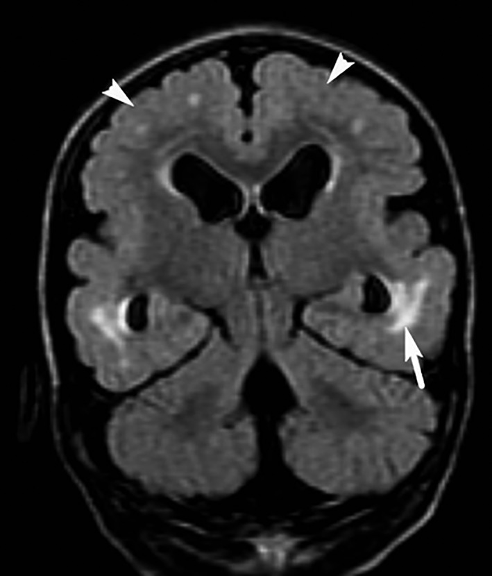
Figure 3.

Brain MRI of younger daughter – FLAIR coronal view, clearly illustrating patches of increased T2 signal (arrow), as well as polymicrogyria and thickened cerebral cortex (arrow heads) with poorly defined boundaries between gray and white matter.
