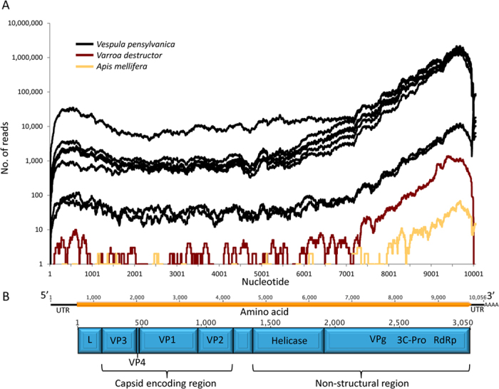
Figure 2.

(A) Moku virus genome coverage from Illumina Hi-seq data for samples collected in Hawaii. V. pensylvanica are shown in black, Varroa in red and honey bees in yellow. Three different honey bee Illumina runs were pooled together for the honey bee data. (B) Organisation of the 10,056 nucleotides Moku virus genome (black line) coding for a 3050-aa polyprotein (orange box) and the predicted polyprotein coding regions are shown in blue.
