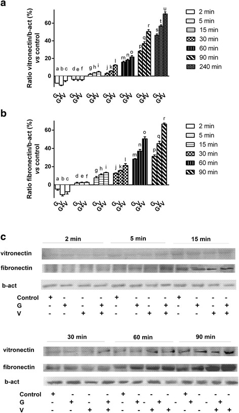
Fig. 2.

Western blot and densitometric analysis of vitronectin and fibronectin. G = Grisù®; V = vitD3; G + V = Grisù® combined with vitD3. The ratio reports a mean ± (SD) (%) of 5 biological replicates normalized to control values (line 0 %). In a densitometric analysis of vitronectin normalized to control values during time (from 2 to 240 min). a, b, c, d, e, f, h, i, j, k, l, m, n, o, p, q, r, s, t, u p < 0.05 vs control; a, b p < 0.05 vs c; e p < 0.05 vs f; i p < 0.05 vs g; j, k p < 0.05 vs l; m, n, p < 0.05 vs o; p, q p < 0.05 vs r; s, t p < 0.05 vs u. In b densitometric analysis of fibronectin normalized to control values during time (from 2 to 90 min). d, e, f, g, h, i, j, k, l, m, n, o, p, q, r p < 0.05 vs control; g, h p < 0.05 vs i; j, k p < 0.05 vs l; m, n p < 0.05 vs o; p, q p < 0.05 vs r. In c an example of Western blot of protein extracts analyzed by immunoblotting with specific antibodies against the indicated proteins
