Figure 4.

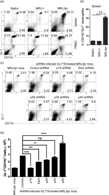
Interleukin (IL)‐39‐deficient GL7+ B cells could not induce neutrophils in lupus‐prone mice. (a,b) CD11b+Gr‐1+ neutrophils from spleen and peripheral blood mononuclear cells (PBMC) of 8‐month‐old female BALB/C, non‐lupus‐prone Murphy Roths large (MRL)+ and lupus‐prone MRL/lpr mice (six mice per group) were analysed by fluorescence activated cell sorter (FACS). The percentages of CD11b+Gr‐1+ neutrophils in the spleen and PBMC (a) and statistical analysis of the percentages in the spleen (b) are shown. (c,d) GL7+ B cells from 8‐month‐old female lupus‐prone MRL/lpr mice were sorted by FACS and infected with control shRNA or IL‐12 family subunits p28, p35 or p40, p19 or Epstein–Barr virus‐induced 3 (Ebi3)‐specific shRNA. On day 1 after infection, 5 × 10⁶ control, p28, p35, p40, p19 and Ebi3‐specific shRNA‐infected GL7+B220+ B cells per mouse were injected intravenously (i.v.) into 8‐week‐old female lupus‐prone MRL/lpr mice (six mice per group). Age‐matched MRL/+ mice and control shRNA‐infected GL7+ B cells transferred group were used as non‐lupus‐prone mice and control shRNA, respectively. On day 14 after cell transfer, splenocytes are analysed by FACS. The percentages (c) and the absolute numbers (d) of CD11b+Gr‐1+ neutrophils per spleen are shown. We used two‐tailed Student's t‐test to analyse the difference between each of p28, p35 or p40, p19 or Ebi3‐specific shRNA‐infected GL7+ B cells transfer group and control shRNA‐infected GL7+ B cells transfer group. (b,d) Data are shown as mean ± standard error of the mean (s.e.m.) (n = 6) from one experiment representative of two other similar experiments. **P < 0·01; ***P < 0·001; ****P < 0·0001. One‐way analysis of variance (anova) plus Dunnett's multiple comparison test: compare all columns versus control column. Error bars, s.e.m.
