Fig. 1.

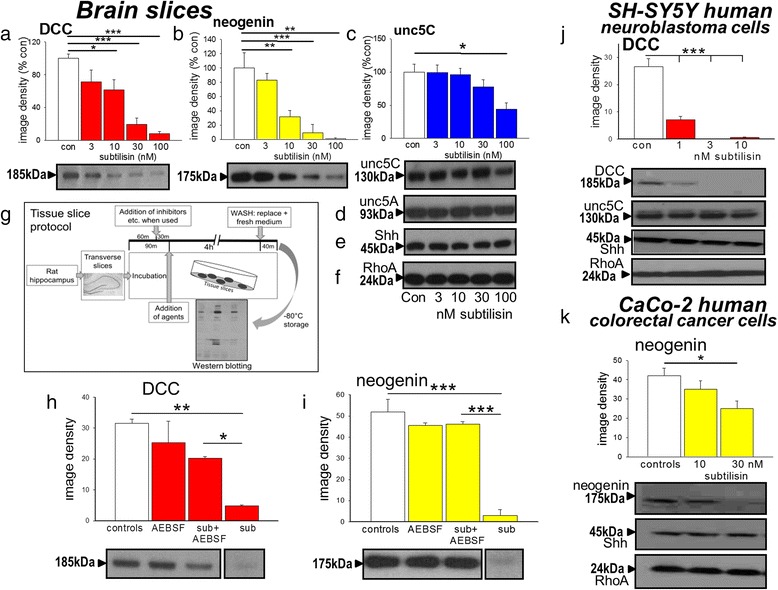
Effects of subtilisin on protein expression. Protein expression in extracts of brain slices is summarised as image densities (arbitrary units) of Western blots quantified using Image J for the effects of subtilisin on (a) DCC (b) neogenin, (c) Unc-5C, (d) Unc-5A, (e) Shh and (f) RhoA expression. Sample blots are shown below each chart (a-c), which illustrate the concentration-dependent effects of subtilisin and the selectivity of its effects. Scheme (g) is a graphic summary of the experimental protocol for these and subsequent experiments. Panels (h) and (i) summarise the blockade by AEBSF (100 μM, 4 h) of the depletion of DCC and neogenin by subtilisin (sub, 30nM), confirming the role of serine protease activity. Panel (j) summarises the effects of subtilisin (1, 3 and 10nM) on the expression of DCC (chart and blot) and on unc-5C, Shh and RhoA (blots) after 7 days in cultures of SH-SY5Y cells. Human colorectal cancer CaCo-2 cells showed a similar susceptibility with reduced expression of neogenin and unc-5C (k) but no change in RhoA or Shh after 7days in subtilisin at 10 or 30nM. Bars represent mean ± s.e.mean (n = 4). *P < 0.05; **P < 0.01; ***P < 0.001 relative to the control bar
