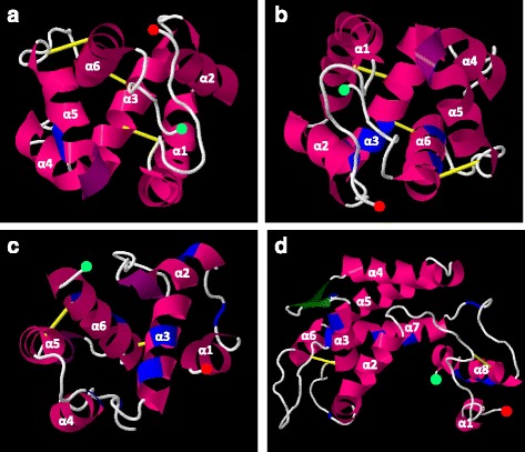
Fig. 4.

Cartoon representation of hypothetical positions of the positively selected sites in Anastrepha OBPs, based in their predicted 3D structures. α-helices are shown in pink, β-sheets in green, loops in white, disulphide bonds in yellow and positively selected sites in blue. N- and C-terminus residues are shown with red and light green circles, respectively. a) AoblOBP56h-1 and AfraOBP56h-1 representation; b) AoblOBP56h-2 and AfraOBP56h-2 representation; c) AoblOBP57c and AfraOBP57c representation; d) AoblOBP50a, AfraOBP50a-1 and AfraOBP50a-2 representation
